Noticing changes in your skin can be unsettling. You might discover a rough patch that feels like sandpaper or a spot that simply refuses to heal. For many, these small, scaly lesions are the first indication of long-term sun exposure catching up with them.
These patches are known as actinic keratoses (AKs), also frequently called solar keratoses. While the term might sound intimidating, understanding what they are is the first step towards effective management.
They are extremely common, particularly among those who have spent significant time outdoors without adequate protection. While they are not cancer, they are considered precancerous. This means that if left untreated, there is a small risk they could develop into a type of skin cancer known as squamous cell carcinoma.
The good news is that these lesions are highly treatable. By learning to recognise actinic keratosis symptoms early, you can prioritise your skin health and prevent more serious complications down the line.
Here is everything you need to know about identifying, treating, and preventing actinic keratosis.
What is actinic keratosis?
Actinic keratosis is a rough, scaly patch on the skin that develops from years of exposure to the sun. It is essentially evidence of damage to the skin cells (keratinocytes) caused by ultraviolet (UV) radiation.
Because the damage is cumulative, these lesions often appear on areas of the body most exposed to the sun, such as the face, lips, ears, back of the hands, forearms, scalp, and neck.
Why is it called “precancerous”?
Medical professionals classify AKs as precancerous because they have the potential to turn into skin cancer. However, this does not mean every patch will become malignant.
According to the NHS, while most actinic keratoses do not turn into cancer, there is a risk they can transform into squamous cell carcinoma (SCC) if left untreated. Because it is clinically difficult to predict exactly which lesions will progress, dermatologists generally recommend treating them all.
Who gets them?
Anyone can develop these lesions, but certain factors increase your likelihood. You are at higher risk if you:
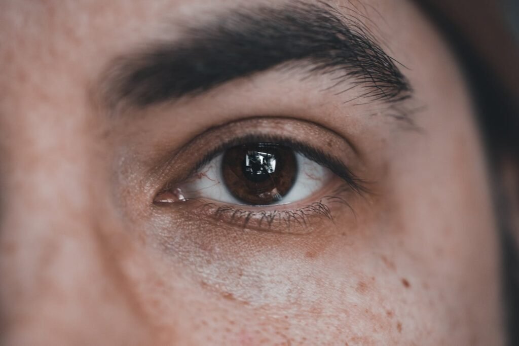
- Have fair skin, blue or green eyes, and red or blonde hair.
- Have a history of intense or frequent sun exposure or sunburns.
- Are over the age of 40.
- Have a weakened immune system due to chemotherapy, leukaemia, AIDS, or organ transplant medications.
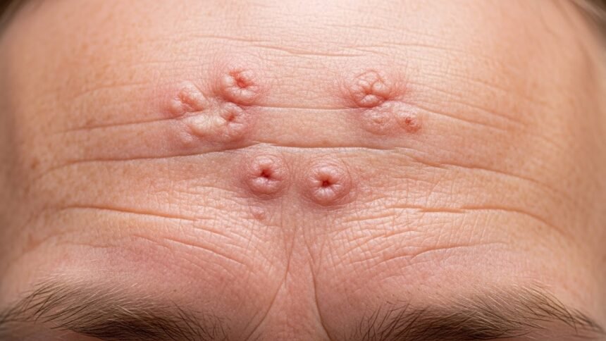
Recognising the signs: Actinic keratosis symptoms
Identifying an AK can be tricky because they vary significantly in appearance. Sometimes, you might feel them before you actually see them. Understanding the nuance of actinic keratosis symptoms is vital for early detection.
Texture and Sensation
The most distinctive feature of an AK is often its texture. Many people discover them by running their hands over their skin rather than by looking in the mirror.
- Sandpaper feel: The lesion often feels rough, dry, and scaly, much like a piece of sandpaper.
- Tenderness: In some cases, the area may feel sensitive or prickly.
- Itching or burning: You might experience a mild stinging sensation, though many AKs are painless.
If you have a rough patch that persists despite moisturising, it is worth investigating further.
Visual Appearance
Visually, actinic keratosis can present in several ways. The lesions usually start small and grow slowly.
- Colour: They can range from skin-coloured to pink, red, or brown. On darker skin tones, they may appear grey or hyperpigmented.
- Size: Most range from a few millimetres to a couple of centimetres in diameter.
- Shape: They can be flat or slightly raised. Sometimes they develop a hard, wart-like surface.
- Crusting: You may notice a yellow or white crust that scales off but returns later.
For a visual guide on what to look for, reliable resources like DermNet NZ offer extensive image libraries of these skin conditions.
The “Horn” Variation
In rare instances, an actinic keratosis can grow rapidly upward, resembling a small animal horn. This is known as a cutaneous horn. The base of this horn may contain squamous cell carcinoma, so immediate medical attention is required.
Common Locations
Because UV radiation is the primary cause, look for symptoms in high-exposure areas:
- The Scalp: Especially common in men with thinning hair.
- The Ears: Look at the rims and the tops of the ears.
- The Face: Forehead, nose, and cheeks are prime locations.
- The Lips: This variation is called actinic cheilitis and presents as cracked, dry, or scaly lips.
Causes and Diagnosis
Understanding the root cause helps in prevention. The primary culprit is chronic exposure to UV light—either from the sun or tanning beds.
How UV damage works
When UV light hits your skin, it damages the DNA within your skin cells. The body can repair some of this damage, but repeated exposure overwhelms the body’s repair mechanisms. Over time, these damaged cells mutate and reproduce, forming the scaly patches characteristic of AK.
According to Cancer Research UK, getting sunburnt just once every two years can triple your risk of malignant melanoma, and cumulative exposure is directly linked to non-melanoma skin cancers and AKs.
Diagnosing the condition
If you spot a suspicious lesion, you should see a GP. Diagnosis is usually straightforward:
- Visual Exam: A doctor will examine your skin, likely using a magnifying device called a dermatoscope.
- Biopsy: If the doctor suspects the lesion has already progressed to cancer, they may perform a skin biopsy. This involves removing a small sample of the tissue for analysis in a laboratory.
Early diagnosis is key. Healthline notes that treating these lesions early is the most effective way to prevent the development of skin cancer.
Comparison: Is it Actinic Keratosis or something else?
It can be difficult to distinguish AKs from other common skin conditions. The table below outlines key differences to help you understand what you might be seeing.
| Feature | Actinic Keratosis | Seborrhoeic Keratosis | Eczema/Dry Skin | Squamous Cell Carcinoma |
|---|---|---|---|---|
| Cause | UV Damage | Ageing / Genetics | Irritation / Allergies | UV Damage / Evolution of AK |
| Texture | Rough, sandpaper-like | Waxy, “stuck-on” appearance | Dry, flaky | Thick, crusty, ulcerated |
| Sensation | Prickly, tender, or painless | Usually painless, can itch | Itchy | Tender, painful, may bleed |
| Location | Sun-exposed areas | Back, chest, face, neck | Elbows, knees, hands | Sun-exposed areas |
| Cancer Risk | Precancerous | Benign (Non-cancerous) | None | Malignant (Cancerous) |
Treatment Options
Treatment varies depending on the number of lesions and their appearance. Your dermatologist or GP will suggest the best course of action.
Topical Treatments
For widespread lesions, creams and gels are often prescribed. These are known as “field treatments” because they treat an area of skin rather than just one spot.
- 5-fluorouracil (5-FU): A chemotherapy cream applied to the skin that destroys precancerous cells.
- Imiquimod: A cream that stimulates your immune system to fight the abnormal cells.
- Diclofenac gel: A non-steroidal anti-inflammatory drug that is gentler but may take longer to work.
Procedural Treatments
For individual or thicker lesions, physical removal is often preferred.
- Cryotherapy: The most common treatment involving freezing the lesion with liquid nitrogen. The British Association of Dermatologists explains that this causes the lesion to blister and fall off after a few days.
- Curettage and Cautery: Scraping the lesion away and using heat to seal the wound.
- Photodynamic Therapy (PDT): A special cream is applied to the skin, which is then exposed to a specific light aimed at killing the abnormal cells.
For more detailed guidance on treatments, you can refer to the guidelines provided by the Mayo Clinic, which outlines the recovery times associated with each method.
Prevention Tips
Prevention is always better than cure. Even if you have already developed AKs, protecting your skin now can prevent new ones from forming.
- Limit Sun Exposure: Avoid the sun when it is strongest, typically between 11 am and 3 pm in the UK.
- Wear Protective Clothing: Broad-brimmed hats, long sleeves, and sunglasses provide a physical barrier.
- Use Sunscreen: Apply a broad-spectrum sunscreen with an SPF of at least 30 (preferably 50). The Skin Cancer Foundation emphasises applying sunscreen every day, regardless of the weather.
- Avoid Tanning Beds: These emit concentrated UV radiation that significantly accelerates skin damage.
- Regular Self-Checks: Examine your skin monthly for new growths or changes in existing moles and freckles.
For comprehensive advice on sun safety standards, GOV.UK provides resources on how to stay safe during hot weather.
Frequently Asked Questions
Is actinic keratosis contagious?
No, actinic keratosis is not contagious. You cannot catch it from someone else, nor can you spread it to other parts of your body through contact. It is strictly the result of UV damage.
Will actinic keratosis go away on its own?
Occasionally, an AK may disappear on its own, especially if you reduce sun exposure. However, they often return. Medical intervention is usually required to ensure they are fully resolved.
Can diet affect actinic keratosis?
While no specific diet cures AK, a diet rich in antioxidants helps support overall skin health. Some studies suggest that low-fat diets may reduce the development of new lesions, though more research is needed.
What happens if I scratch it off?
It is tempting to scratch off the crusty skin, but this is not recommended. It can lead to infection, scarring, and will not remove the damaged cells deeper in the skin. The lesion will almost certainly grow back.
The Bottom Line
Actinic keratosis is a warning sign from your body that your skin has sustained significant sun damage. While the word “precancerous” is frightening, the prognosis for those who seek treatment is excellent.
By staying vigilant about actinic keratosis symptoms—specifically looking for those rough, sandpaper-like patches—you can catch these lesions early. Treatment is generally quick, effective, and minimally invasive.
If you notice a spot that is changing, bleeding, or simply not healing, book an appointment with your GP or a dermatologist. Your skin is your body’s largest organ, and protecting it is an investment in your long-term health.
For further reading on managing skin conditions, reliable sources like Johns Hopkins Medicine offer in-depth patient education materials.